
ファイル:DTI-sagittal-fibers.jpg
このプレビューのサイズ: 643 × 600 ピクセル。 その他の解像度: 257 × 240 ピクセル | 515 × 480 ピクセル | 1,021 × 952 ピクセル。
元のファイル (1,021 × 952 ピクセル、ファイルサイズ: 294キロバイト、MIME タイプ: image/jpeg)
|
概要
| 解説 |
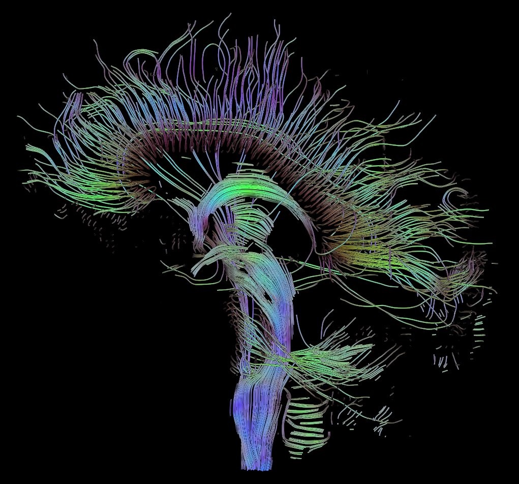
English: Visualization of a DTI measurement of a human brain. Depicted are reconstructed fiber tracts that run through the mid-sagittal plane. Especially prominent are the U-shaped fibers that connect the two hemispheres through the corpus callosum (the fibers come out of the image plane and consequently bend towards the top) and the fiber tracts that descend toward the spine (blue, within the image plane)
Français : Visualisation d'une mesure DTI d'un cerveau humain. Ce qui est représenté sont des faisceaux de fibres reconstruits qui traversent le plan demi-sagittal. On observe les fibres en U qui connectent les deux hémisphères à travers le corps calleux, qui sont particulièrement importantes (les fibres sortent du plan de l'image et par conséquent se courber vers le haut) ainsi que les faisceaux de fibres qui descendent vers la colonne vertébrale (bleu, dans le plan de l'image)
Deutsch: Traktographie-Verfahren rekonstruieren aus den Messdaten der Diffusions-Tensor-Bildgebung den anzunehmenden Verlauf größerer Nervenbahnen. Hier dargestellt sind die Ergebnisse für ein menschliches Gehirn; um die Übersichtlichkeit zu wahren, beschränkt sich die Abbildung auf Bahnen, die die Medianebene schneiden. Insbesondere sind dies die U-förmigen Faserbündel, die die beiden Hirnhälften verbinden (sie durchstoßen die Bildebene und sind nach oben gebogen) sowie die Faserbündel, die zum Rückenmark ziehen (blau dargestellt, liegen innerhalb der Bildebene) |
| 日付 | |
| 原典 | 投稿者自身による著作物 |
| 作者 | Thomas Schultz |
| 許可 (ファイルの再利用) |
Rendering is own work, using a modified version of the BioTensor application developed at the University of Utah. The dataset is courtesy of Gordon Kindlmann at the Scientific Computing and Imaging Institute, University of Utah, and Andrew Alexander, W.M. Keck Laboratory for Functional Brain Imaging and Behaviour, University of Wisconsin, Madison. It is publicly available from [1] |
ライセンス
この作品の著作権者である私は、この作品を以下のライセンスで提供します。

|
この文書は、フリーソフトウェア財団発行のGNUフリー文書利用許諾書 (GNU Free Documentation License) 1.2またはそれ以降のバージョンの規約に基づき、複製や再配布、改変が許可されます。不可変更部分、表紙、背表紙はありません。このライセンスの複製は、GNUフリー文書利用許諾書という章に含まれています。 |
| このファイルはクリエイティブ・コモンズ 表示-継承 3.0 非移植ライセンスのもとに利用を許諾されています。 | ||
| ||
| このライセンスのテンプレートは、GFDLのライセンス・アップデートによりこのファイルに追加されたものです。 |
- あなたは以下の条件に従う場合に限り、自由に
- 共有 – 本作品を複製、頒布、展示、実演できます。
- 再構成 – 二次的著作物を作成できます。
- あなたの従うべき条件は以下の通りです。
- 表示 – あなたは適切なクレジットを表示し、ライセンスへのリンクを提供し、変更があったらその旨を示さなければなりません。これらは合理的であればどのような方法で行っても構いませんが、許諾者があなたやあなたの利用行為を支持していると示唆するような方法は除きます。
- 継承 – もしあなたがこの作品をリミックスしたり、改変したり、加工した場合には、あなたはあなたの貢献部分を元の作品とこれと同一または互換性があるライセンスの下に頒布しなければなりません。
あなたは上記のライセンスから、どれか一つ以上を選択できます。
ファイルの履歴
過去の版のファイルを表示するには、その版の日時をクリックしてください。
| 日付と時刻 | サムネイル | 寸法 | 利用者 | コメント | |
|---|---|---|---|---|---|
| 現在の版 | 2017年10月13日 (金) 10:42 | | 1,021 × 952 (294キロバイト) | Mikael Häggström | Minor crop of black areas at the top and bottom |
| 2006年9月22日 (金) 16:22 |  | 1,021 × 1,125 (203キロバイト) | Thomas Schultz | {{Information |Description=Visualization of a DTI measurement of a human brain. Depicted are reconstructed fiber tracts that run through the mid-sagittal plane. Especially prominent are the U-shaped fibers that connect the two hemispheres through the corp |
リンク
この画像にリンクしているページはありません。
グローバルなファイル使用状況
以下に挙げる他のウィキがこの画像を使っています:
- af.wikipedia.org での使用状況
- ar.wikipedia.org での使用状況
- az.wikiquote.org での使用状況
- bn.wikipedia.org での使用状況
- cs.wikipedia.org での使用状況
- de.wikipedia.org での使用状況
- Autismus
- Computergrafik
- Bipolare Störung
- Portal:Informatik/Exzellente Artikel
- Portal:Geist und Gehirn/Artikel des Monats
- Diffusions-Tensor-Bildgebung
- Wikipedia:Kandidaten für exzellente Bilder/Archiv2006/17
- Datei:DTI-sagittal-fibers.jpg
- Wikipedia:Exzellente Bilder/Naturwissenschaften
- Portal:Physik/Artikel des Monats 2024-03
- Wikipedia:Exzellente Bilder/Kleine Bilder
- en.wikipedia.org での使用状況
- Neurolinguistics
- Tractography
- Portal:Medicine
- User talk:Spikebrennan
- User:Spikebrennan
- Diffusion MRI
- Wikipedia:WikiProject Neuroscience
- Portal:Psychology/Selected article
- Wikipedia:Featured pictures/Sciences/Biology
- Portal:Psychology/Selected article/7
- Wikipedia:Featured pictures thumbs/08
- Wikipedia:Featured picture candidates/DTI-sagittal-fibers.jpg
- Wikipedia:Wikipedia Signpost/2007-11-05/Features and admins
- Wikipedia:Featured picture candidates/November-2007
- Wikipedia:Picture of the day/March 2008
- Connectome
- Template:POTD/2008-03-10
- User talk:Thomas Schultz
- Wikipedia:Wikipedia Signpost/2007-11-05/SPV
- Biological data visualization
- Wikipedia:WikiProject Medicine/Recognized content
- Wikipedia:WikiProject Molecular Biology/Biophysics
- User:Wouterstomp/test
- Wikipedia:WikiProject Anatomy/Resources
- Wikipedia:WikiProject Anatomy/Recognized content
- Wikipedia talk:WikiProject Anatomy/Archive 9
- Portal:Medicine/Recognized content
- User talk:Rhododendrites/Reconsidering FPC on the English Wikipedia
- User:Hydrogenkitsch
- Wikipedia:Wikipedia Signpost/Single/2007-11-05
- en.wikibooks.org での使用状況
このファイルのグローバル使用状況を表示する。